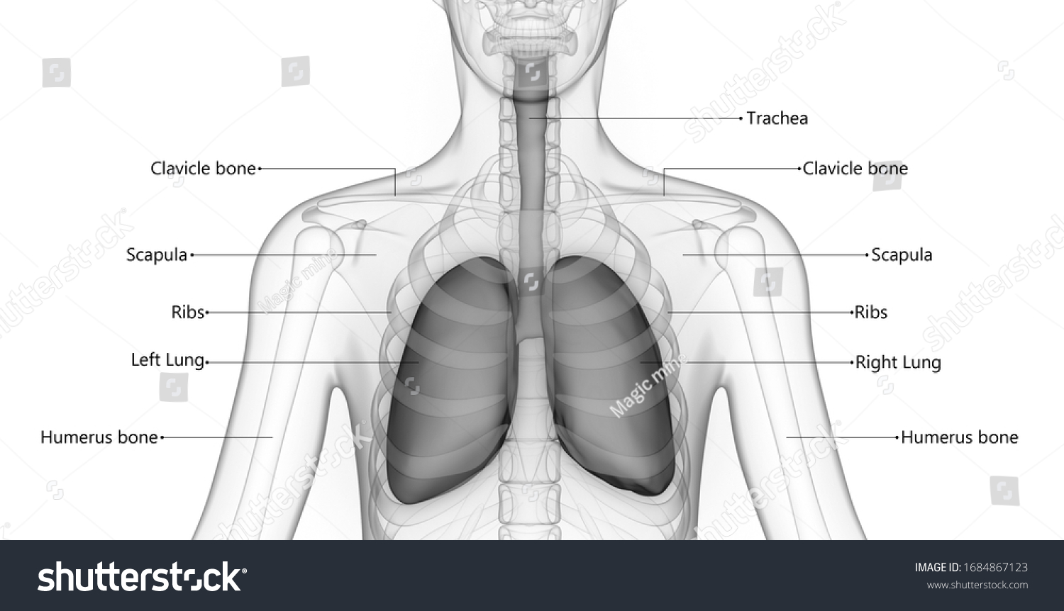
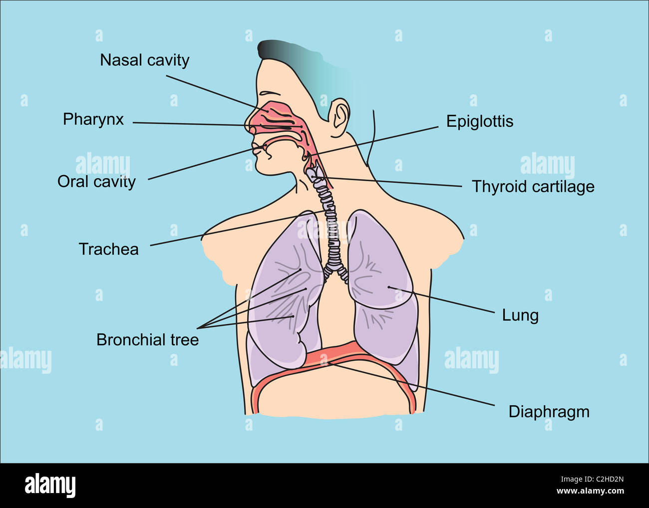
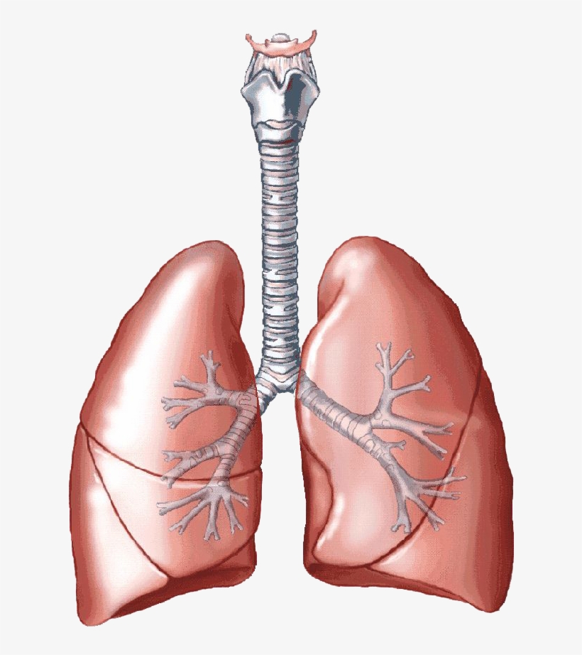
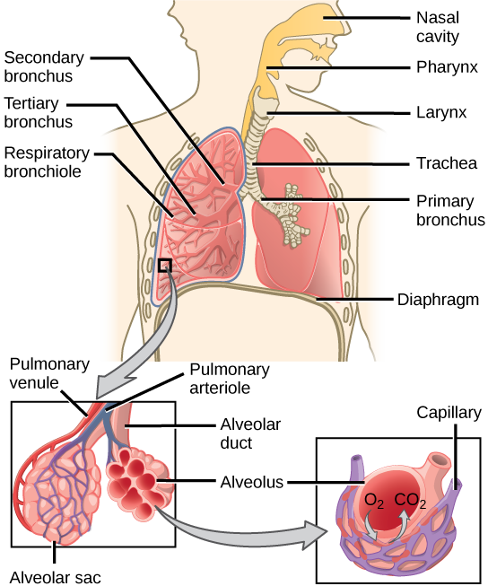
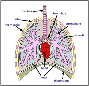
39 diagram of the lungs with labels
› circulatory-system-diagramCirculatory System Diagram - Cardiovascular System and Blood ... They may come with or without labels. Common circulatory system diagrams show pulmonary circulation, coronary circulation, systematic circulation, veins, arteries, or a combination. The systemic circulation system is the most commonly illustrated of the systems that make up the circulatory system as it is the largest. en.wikipedia.org › wiki › CigaretteCigarette - Wikipedia The widespread smoking of cigarettes in the Western world is largely a 20th-century phenomenon. At the start of the 20th century, the per capita By 2000, consumption had fallen to 2,092 per capita, corresponding to about 30% of men and 22% of women smoking more than 100 cigarettes per year, and by 2006 per capita consumption had declined to 1,691; implying that about 21% of the population ...
8,956 Lung diagram Images, Stock Photos & Vectors | Shutterstock 8,956 lung diagram stock photos, vectors, and illustrations are available royalty-free. See lung diagram stock video clips Image type Orientation Sort by Popular Healthcare and Medical Anatomy Recreation/Fitness lung respiratory system medicine pulmonary alveolus organ breathing human body Next of 90
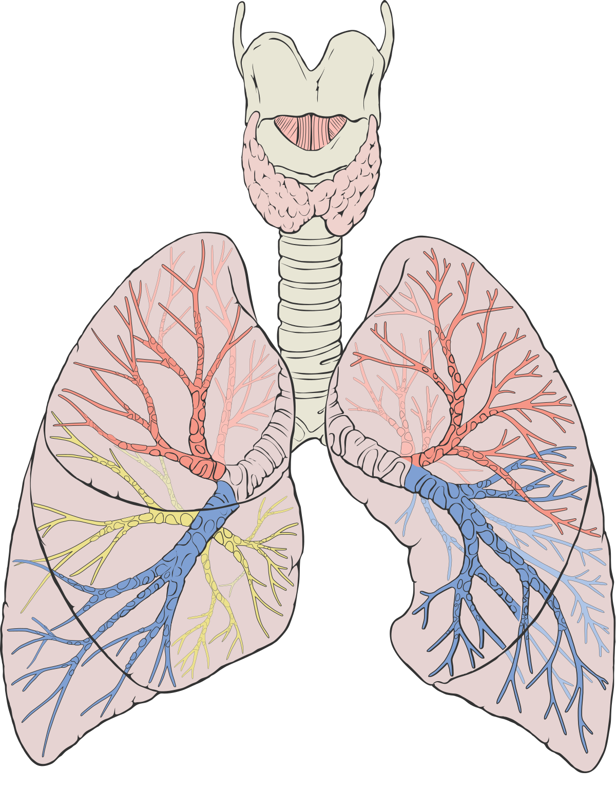
Diagram of the lungs with labels
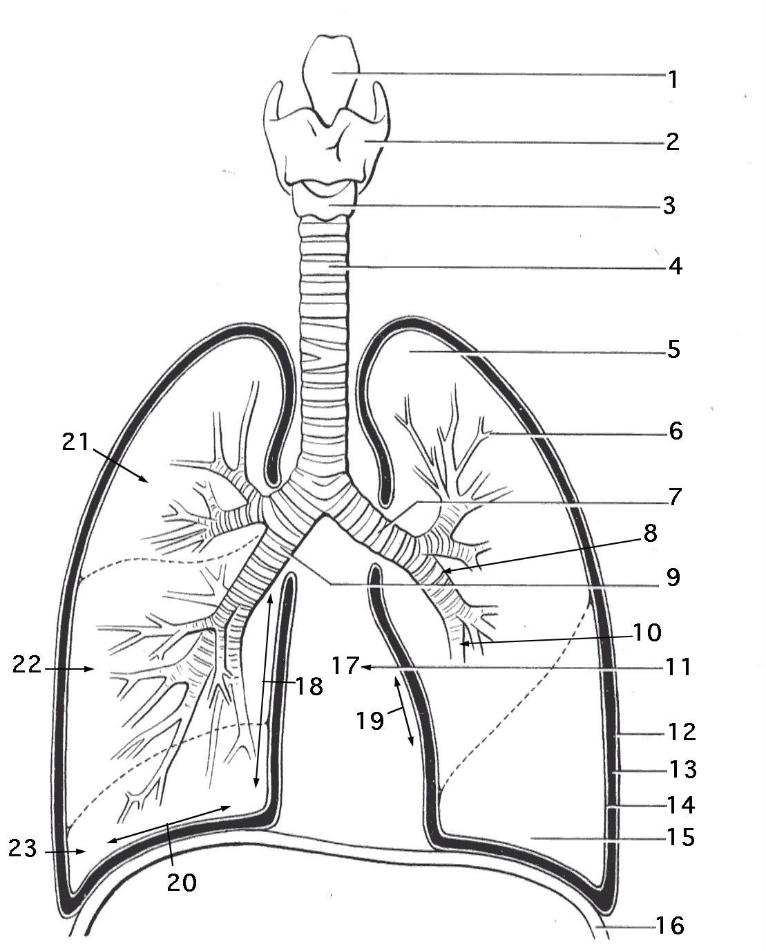
Respiratory system quizzes and labeled diagrams | Kenhub We've got plenty of respiratory system quizzes containing questions on everything from the lymphatics of the lungs to the bronchi, bronchioles and alveoli. Try one right now! Check out some more of our top picks about the respiratory system below. Trachea Explore study unit. Bronchial tree and alveoli Explore study unit. › createJoin LiveJournal Password requirements: 6 to 30 characters long; ASCII characters only (characters found on a standard US keyboard); must contain at least 4 different symbols; Labeled diagram of the lungs/respiratory system. - SERC View Original Image at Full Size. Labeled diagram of the lungs/respiratory system. Image 37789 is a 1125 by 1408 pixel PNG Uploaded: Jan10 14. Last Modified: 2014-01-10 12:15:34
Diagram of the lungs with labels. Lung Anatomy, Function, and Diagrams - Healthline The lungs begin at the bottom of your trachea (windpipe). The trachea is a tube that carries the air in and out of your lungs. Each lung has a tube called a bronchus that connects to the trachea.... Fully Labelled Diagram Alveolus Lungs Showing Stock ... - Shutterstock High Usage score High usage Superstar Shutterstock customers love this asset! Stock Vector ID: 369984683 Fully labelled diagram of the alveolus in the lungs showing gaseous exchange. Vector Formats EPS 1114 × 800 pixels • 3.7 × 2.7 in • DPI 300 • JPG Show more Vector Contributor S Steve Cymro Similar images See all Assets from the same collection byjus.com › biology › human-heartHuman Heart - Anatomy, Functions and Facts about Heart - BYJUS The right ventricle pumps the blood to the lungs for re-oxygenation through the pulmonary arteries. The right semilunar valves close and prevent the blood from flowing back into the heart. Then, the oxygenated blood is received by the left atrium from the lungs via the pulmonary veins. Read on to explore more about the structure of the heart. Anatomy, medical imaging and e-learning for healthcare IMAIOS and selected third parties, use cookies or similar technologies, in particular for audience measurement. Cookies allow us to analyze and store information such as the characteristics of your device as well as certain personal data (e.g., IP addresses, navigation, usage or geolocation data, unique identifiers).
byjus.com › biology › diagram-of-heartHeart Diagram with Labels and Detailed Explanation - BYJUS The diagram of heart is beneficial for Class 10 and 12 and is frequently asked in the examinations. A detailed explanation of the heart along with a well-labelled diagram is given for reference. Well-Labelled Diagram of Heart. The heart is made up of four chambers: The upper two chambers of the heart are called auricles. Parents (for Parents) - Nemours KidsHealth They still put nicotine or chemicals in the body and can damage the lungs. Get the facts. Managing Your Toddler's Behavior. Learn how to encourage good behavior, handle tantrums, and keep your cool when parenting your toddler. Questions and Answers. How can I teach my kids to be smart on social media? › science › answerHuman Body Worksheets - Easy Teacher Worksheets In the diagram to the left, provide the labels for the structures involved in the reflex act when a person steps on a tack and jerks their leg away. Brain Anatomy Provide the labels for the diagram on the left below and provide descriptions of the functions of each structure on the blank lines. How to draw and label a lung | step by step tutorial - YouTube A beautiful drawing of Lung. And it will teach you to draw the lung very easily. Watch the video and please be kind enough to thumbs up my videos. And I will...
01. Characteristics and Classification of Living Organisms 1. Draw an animal cell and include the following labels: nucleus, cell membrane, cytoplasm, rough endoplasmic reticulum, ribosomes 2. Some organelles are only present in certain types of cell, but some are present in all cells. Tick the correct boxes below: Feature Present in all cells Present only in some cells Nucleus ☐ ☐ PDF ANATOMY OF LUNGS - University of Kentucky Lungs are a pair of respiratory organs situated in a thoracic cavity. Right and left lung are separated by the mediastinum. Texture-- Spongy Color - Young - brown Adults -- mottled black due to deposition of carbon particles Weight-Right lung - 600 gms Left lung - 550 gms. Heart Diagram with Labels and Detailed Explanation - BYJUS The diagram of heart is beneficial for Class 10 and 12 and is frequently asked in the examinations. A detailed explanation of the heart along with a well-labelled diagram is given for reference. ... The pulmonary artery, being an exception, carries deoxygenated blood to the lungs for purification. The veins carry impure blood from different ... Diagram Of The Lungs With Labels Labeling Of The Lungs Label The Lungs ... Diagram Of The Lungs With Labels Labeling Of The Lungs Label The Lungs Diagram Diagram Of Lungs With. By admin Apr 15, 2019. Share this page . Post navigation. Lung Lobectomy: What you need to know . By admin. Related Post. Leave a Reply Cancel reply. You must be logged in to post a comment.
MASTERING AP: CHAPTER 1 Flashcards | Quizlet B) The extremely thin tissue (simple squamous epithelium) of the lungs allows for the quick diffusion of respiratory gases into and out of the body. C) The direction of blood flow through the heart is directed by one way valves. D) The innermost lining of the lungs is composed primarily of a thin tissue called simple squamous epithelium.
Respiratory System Anatomy, Diagram & Function | Healthline Respiratory. The respiratory system, which includes air passages, pulmonary vessels, the lungs, and breathing muscles, aids the body in the exchange of gases between the air and blood, and between ...
Data Collection for Machine Learning: The Complete Guide Sep 28, 2021 · The arrows on the diagram show the most important and frequent dependencies between the phases, while the outer circle symbolizes the very nature of Data Mining in general. Apart from the machine learning application, CRISP-DM has been used widely in various research projects, like medical data analysis, evaluation of heating and air ...
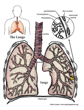
Labeled Diagram of the Human Lungs - Bodytomy Given below is a labeled diagram of the human lungs followed by a brief account of the different parts of the lungs and their functions. Each lung is enclosed inside a sac called pleura, which is a double-membrane structure formed by a smooth membrane called serous membrane.
Lungs Diagram Labeled Illustrations, Royalty-Free Vector Graphics ... Choose from Lungs Diagram Labeled stock illustrations from iStock. Find high-quality royalty-free vector images that you won't find anywhere else.
en.wikipedia.org › wiki › Human_eyeHuman eye - Wikipedia Diagram of a human eye (horizontal section of the right eye) 1. Lens, 2. Zonule of Zinn or Ciliary zonule, 3. Posterior chamber and 4. Anterior chamber with 5. Aqueous humour flow; 6. Pupil, 7. Corneosclera or Fibrous tunic with 8. Cornea, 9. Trabecular meshwork and Schlemm's canal. 10. Corneal limbus and 11. Sclera; 12. Conjunctiva, 13. Uvea ...
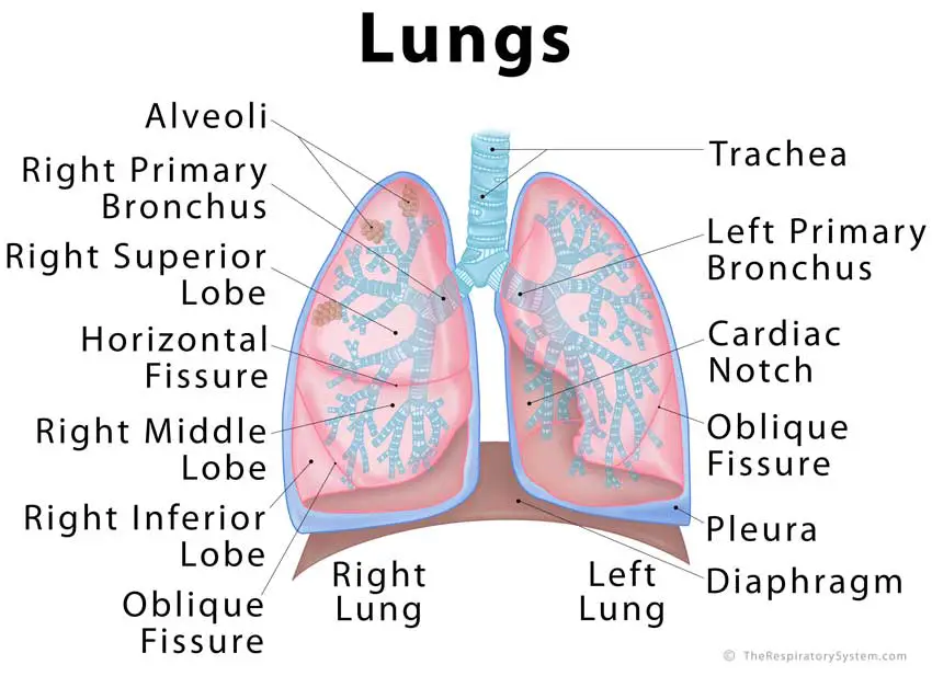
Lung Diagram Labeled | EdrawMax Template in the following lung labeled diagram, we have shown thyroid cartilage, cricoid cartilage, tracheal cartilage, apex, left upper lobe, hilum, left bronchus, oblique fissure, bronchioles, left lower lobe, base of lung, cardiac notch, right lower lobe, oblique fissure, right middle lobe, horizontal fissure, right bronchus, right upper lobe, and …
Consumer Updates | FDA - U.S. Food and Drug Administration Jul 28, 2022 · The .gov means it’s official. Federal government websites often end in .gov or .mil. Before sharing sensitive information, make sure you're on a federal government site.
Label the lung diagram Diagram | Quizlet Start studying Label the lung diagram. Learn vocabulary, terms, and more with flashcards, games, and other study tools.
Label Lungs Diagram Printout - Enchanted Learning bronchial tree: the system of airways within the lungs, which bring air from the trachea to the lung's tiny air sacs (alveoli). cardiac notch: the indentation in the left lung that provides room for the heart. diaphragm: a muscular membrane under the lungs. larynx: a muscular structure at the top of the trachea, containing the vocal cords.
Lungs Diagram - Human Lungs Anatomy - BYJUS The right lung comprises three lobes - inferior, middle and superior lobe that are distinguished by an oblique and deep horizontal fissure. The left lung has two lobes separated by an oblique fissure. The apexes of the lungs expand above the first rib, while both lungs in the thorax rest with their bases on the diaphragm.
Module One- Human Body & Chemistry of Life Flashcards - Quizlet Label the following parts of the diagram depicting negative feedback being used to maintain homeostasis. ... Place each of the labels into the proper category in order to indicate whether they relate to anatomy or physiology. ... pharynx, larynx, trachea, lungs. urinary system-Eliminates nitrogenous wastes from the body. Regulates water ...
Respiratory System Labeling Interactive Quiz - PurposeGames.com lungs. respiratory system. Games by same creator. The States of the Midwest (label all 12 states) ... Label the 6 layers & 4 features of the Sun 10p Image Quiz. The States of the West (label the 13 states) 13p Image Quiz. The Capitals of the Northeastern States Labeling Interactive 9p Image Quiz.
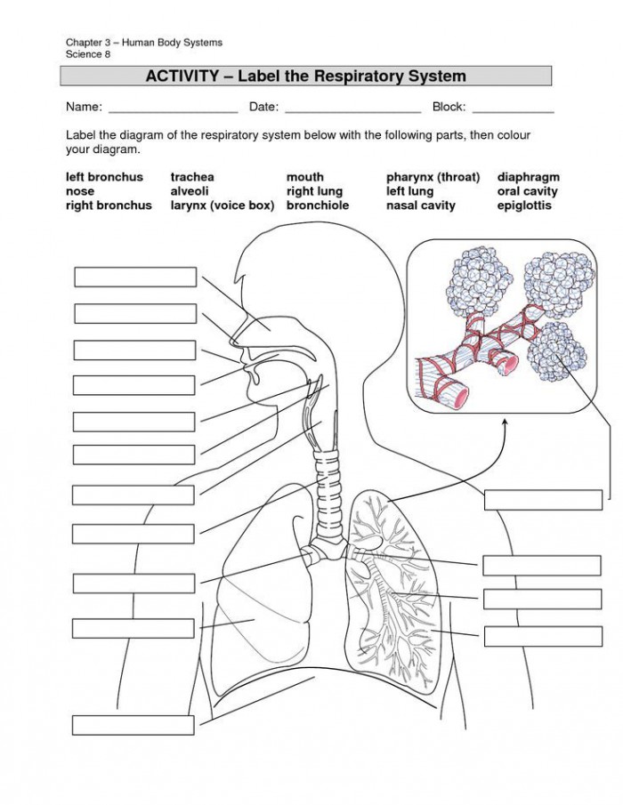
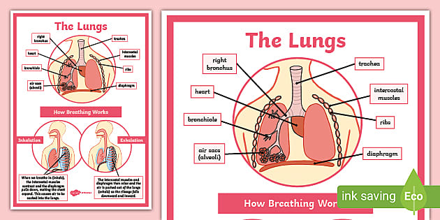
Lung Diagram Labelling Activity | Primary Resources | Twinkl This handy Lung Labelling Worksheet gives your children the opportunity to show how much they've learned about the human lung system. The beautifully hand-drawn illustration shows a lung diagram, labelled with blank spaces where learners can fill in its different components. Encourage your students to work independently and label the parts of the lungs they can see. This teaching resource also ...
Circulatory System Diagram - Cardiovascular System and Blood ... They may come with or without labels. Common circulatory system diagrams show pulmonary circulation, coronary circulation, systematic circulation, veins, arteries, or a combination. The systemic circulation system is the most commonly illustrated of the systems that make up the circulatory system as it is the largest.
Label the Lungs Diagram | Quizlet superior lobe of right lung ... middle lobe of right lung ... inferior lobe of right lung ... superior lobe of left lung ... left main (primary) bronchus ... lobar (secondary) bronchus ... segmental (tertiary) bronchus ... inferior lobe of left lung ... Sets found in the same folder Lab: Heart Model 34 terms enthusiastic_crafter
Diagram Lungs Illustrations & Vectors - Dreamstime Download 2,668 Diagram Lungs Stock Illustrations, Vectors & Clipart for FREE or amazingly low rates! New users enjoy 60% OFF. 194,600,487 stock photos online. ... Labeled diagram with sickness symptoms. Lupus disease vector illustration. Labeled diagram with sickness symptoms, like hair. Diagram of systems in human body. Illustration.
Parts of the Body for Kids: Names & Basic Functions Diagram of Body Parts. External, which means “outside,” describes the body parts that you can see. Take a look at a helpful diagram that labels major external body parts. Download the printable PDF to see it in more detail and print if needed. ... protecting lungs and heart; assisting in arm movement. abdomen. middle of the torso.
Diagram Of The Respiratory System With Labels Pictures, Images ... - iStock In mammals and most other vertebrates, two lungs are located near the backbone on either side of the heart. Vector graphic. Lungs with Alveoli Labeled CG image of woman's chest area showing both lungs in isolation, with magnified view of alveoli air sacs labeled on faded flesh tone and white.
Labeled diagram of the lungs/respiratory system. - SERC View Original Image at Full Size. Labeled diagram of the lungs/respiratory system. Image 37789 is a 1125 by 1408 pixel PNG Uploaded: Jan10 14. Last Modified: 2014-01-10 12:15:34
› createJoin LiveJournal Password requirements: 6 to 30 characters long; ASCII characters only (characters found on a standard US keyboard); must contain at least 4 different symbols;
Respiratory system quizzes and labeled diagrams | Kenhub We've got plenty of respiratory system quizzes containing questions on everything from the lymphatics of the lungs to the bronchi, bronchioles and alveoli. Try one right now! Check out some more of our top picks about the respiratory system below. Trachea Explore study unit. Bronchial tree and alveoli Explore study unit.


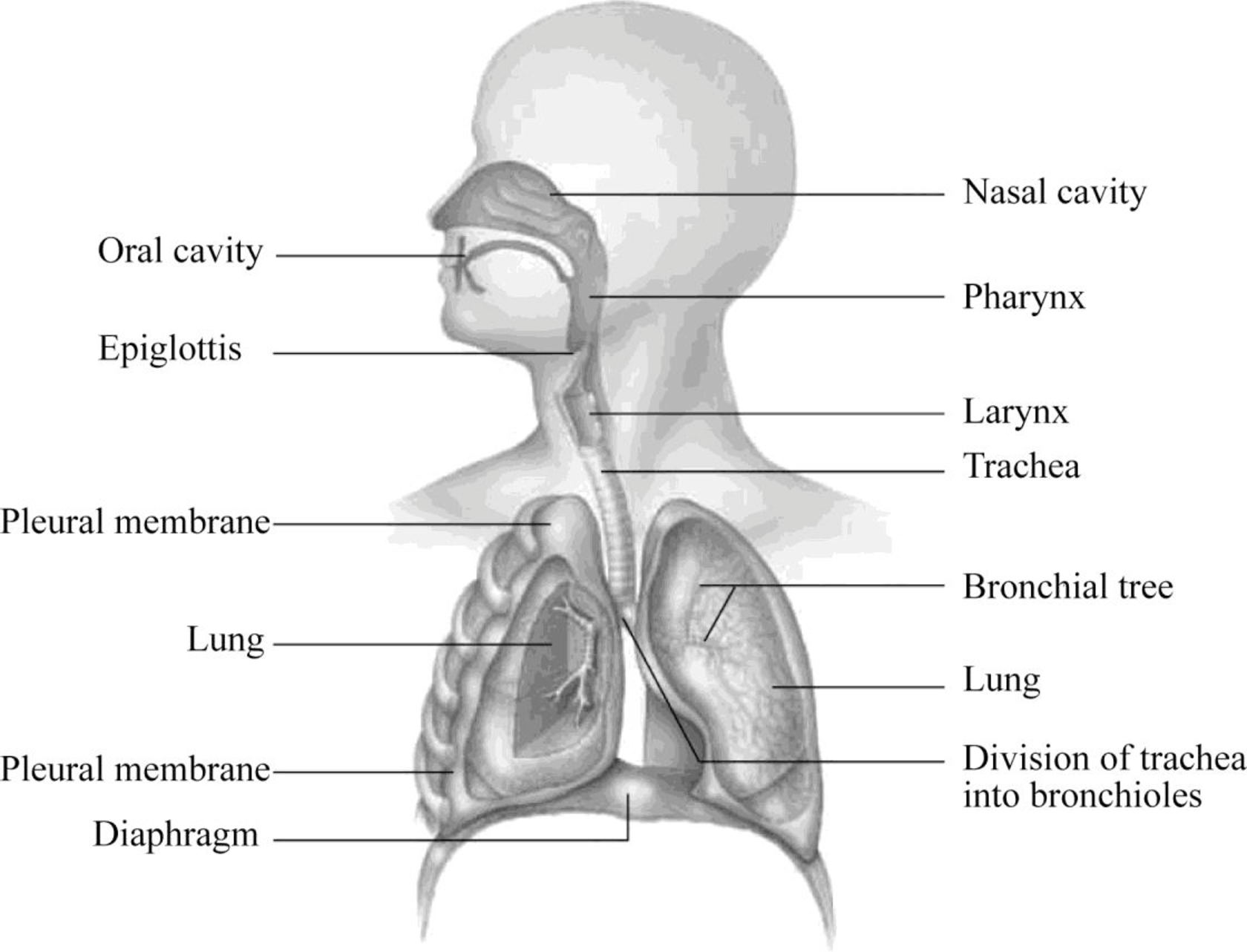

:background_color(FFFFFF):format(jpeg)/images/library/11021/labeled_image_of_the_anatomy_of_respiratory_system.jpg)


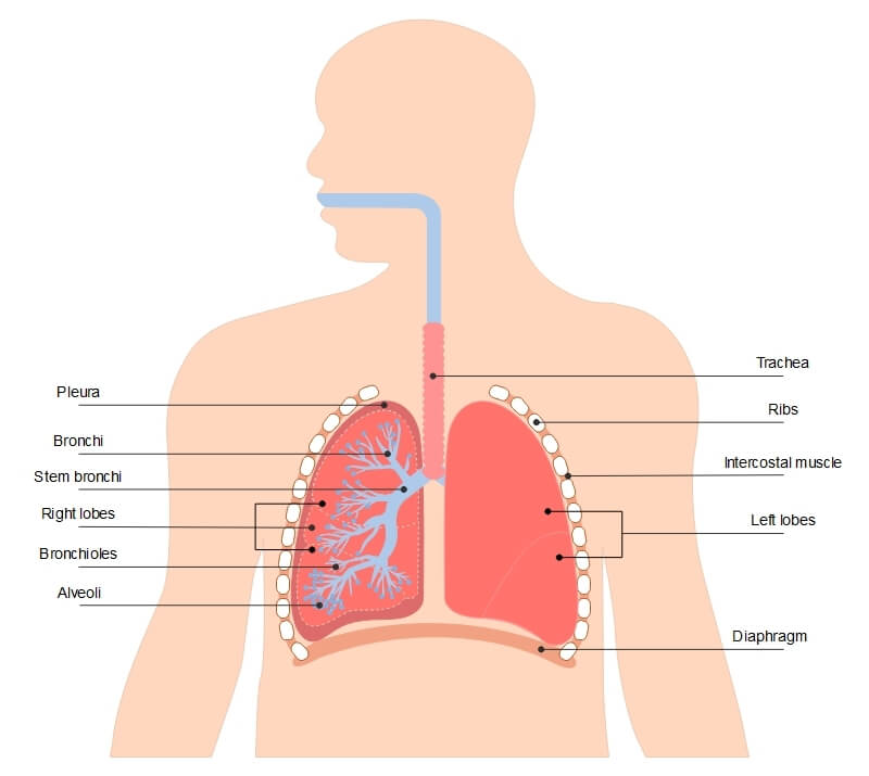






Post a Comment for "39 diagram of the lungs with labels"