42 cell structure with labels
Plant and Animal Cell: Labeled Diagram, Structure, Function - Embibe Double membrane-bound structures found only in the plant cells. 2. This is an autonomous organelle. 3. There are stroma or matrix and grana or stacked discs that are involved in photosynthesis. 4. Grana are the site for photochemical reactions of photosynthesis, while stroma is the site for biochemical reactions of photosynthesis. | automated cell type annotation for scRNA-seq ... The programme uses cell-atlasing and cellular genetics to map cells in the human body combining cutting-edge methodologies and computational approaches. About CellTypist CellTypist was first developed as a platform for exploring tissue adaptation of cell types using scRNA-seq semi-automatic annotations.
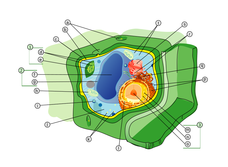
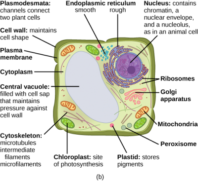
Label the Plant Cell: Level 1 | Worksheet | Education.com In Label the Plant Cell: Level 1, students will use a word bank to label the parts of a cell in a plant cell diagram. To take the learning one step further, have students assign a color to each of the organelles and then color in the diagram. For a broader focus, use this worksheet in conjunction with the Label the Animal Cell: Level 1 ...

Cell structure with labels
en.wikipedia.org › wiki › CarmineCarmine - Wikipedia Carmine (/ ˈ k ɑːr m ə n, ˈ k ɑːr m aɪ n /) – also called cochineal (when it is extracted from the cochineal insect), cochineal extract, crimson lake, or carmine lake – is a pigment of a bright-red color obtained from the aluminium complex derived from carminic acid. Anatomy & Physiology Cell Structure & Function Quiz - Registered Nurse RN This Anatomy & Physiology (A&P) quiz is designed to test your knowledge of the basic cell structure and function. You will be asked questions that pertain to the mitochondria, nucleolus, nuclear membrane, ribosomes, lysosome, and much more. This practice test for the cell function and structure for Anatomy & Physiology, is designed to help you ... Plant Cell: Meaning, Components, Structure, Functions & Parts - Embibe Let us have a detailed look at the plant cell, its structure and the functions of different organelles. Components of a Plant Cell. The small membrane or non-membrane bound structures that are found in the cytoplasm or cellular matrix of a cell that works in a coordinated manner to maintain the homeostasis of a cell are termed as cell organelles.
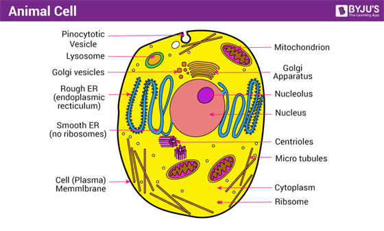
Cell structure with labels. bioinformatics.uconn.edu › single-cell-rnaSingle-cell RNA sequencing (Cell Ranger) | Computational ... The Single Cell 3’ Protocol produces Illumina-ready sequencing libraries. A Single Cell 3’ Library comprises standard Illumina paired-end constructs which begin and end with P5 and P7. The Single Cell 3’ 16 bp 10xTM Barcode and 10 bp randomer is encoded in Read 1, while Read 2 is used to sequence the cDNA fragment. Animal Cell - Structure, Function, Diagram and Types - BYJUS Skin Cells Melanocytes, keratinocytes, Merkel cells and Langerhans cells Muscle Cells Myocyte, Myosatellite cells, Tendon cells, Cardiac muscle cells Blood Cells Leukocytes, erythrocytes, platelet Nerve Cells Schwann cell, glial cells etc Fat Cells Adipocytes Points to Note About Animal Cell The cell is the structural and functional unit of life. › en › productWhat's new in think-cell :: think-cell think-cell 8 greatly expands the slide layout functionality of our software by introducing the pentagon/chevron and textbox as new think-cell elements. Show project steps with accompanying bullet points by creating the basic structure very quickly out of building blocks, and use flexible single-click duplication to add additional steps. Structure of Cell: Definition, Types, Diagram, Functions - Embibe Cells are the fundamental structural and functional unit of all living beings including plants, animals and microorganisms. All living organisms in this universe are made up of cells. We cannot see cells with naked eyes as they are only \ (10\) microns in size whereas human eyes cannot see objects less than \ (100\) microns.
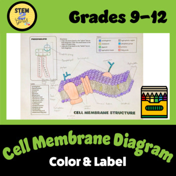
PDF Human Cell Diagram, Parts, Pictures, Structure and Functions The endoplasmic reticulum(ER) is a membranous structure that contains a network of tubules and vesicles. Its structure is such that substances can move through it and be kept in isolation from the rest of the cell until the manufacturing processes conducted within are completed. Human Cell Diagram, Parts, Pictures, Structure and Functions One of the few cells in the human body that lacks almost all organelles are the red blood cells. The main organelles are as follows : cell membrane endoplasmic reticulum Golgi apparatus lysosomes mitochondria nucleus perioxisomes microfilaments and microtubules Diagram of the human cell illustrating the different parts of the cell. Cell Membrane en.wikipedia.org › wiki › History_of_cell_membraneHistory of cell membrane theory - Wikipedia This was the first time the bilayer structure had been universally assigned to all cell membranes as well as organelle membranes. Evolution of the membrane theory. The idea of a semipermeable membrane, a barrier that is permeable to solvent but impermeable to solute molecules was developed at about the same time. Cell: Structure and Functions (With Diagram) - Biology Discussion Eukaryotic Cells: 1. Eukaryotes are sophisticated cells with a well defined nucleus and cell organelles. 2. The cells are comparatively larger in size (10-100 μm). 3. Unicellular to multicellular in nature and evolved ~1 billion years ago. 4. The cell membrane is semipermeable and flexible. 5. These cells reproduce both asexually and sexually.
Eukaryotic Cells- Definition, Characteristics, Structure, & Examples Cell Wall. A cell wall is a rigid structure present outside the plant cell. It is, however, absent in animal cells. It provides shape to the cell and helps in cell-to-cell interaction. It is a protective layer that protects the cell from any injury or pathogen attacks. It is composed of cellulose, hemicellulose, pectins, proteins, etc. Cell Structure | Thermo Fisher Scientific - US Cell structure labels can be a counterstain method to identify the location of specific proteins and targets of interest within the cell. Our cell labels include cell structure specific dyes or antibodies for live-cell or fixed-cell imaging, as well as for flow cytometry. Cell structure protocols Cytoskeleton Structure › books › NBK26880Looking at the Structure of Cells in the Microscope ... A typical animal cell is 10–20 μm in diameter, which is about one-fifth the size of the smallest particle visible to the naked eye. It was not until good light microscopes became available in the early part of the nineteenth century that all plant and animal tissues were discovered to be aggregates of individual cells. Cell Organelles - Types, Structure and their Functions - BYJUS Ribosomes are found in the form of tiny particles in a large number of cells and are mainly composed of 2/3rd of RNA and 1/3rd of protein. They are named as the 70s (found in prokaryotes) or 80s (found in eukaryotes) The letter S refers to the density and the size, known as Svedberg's Unit. Both 70S and 80S ribosomes are composed of two subunits.
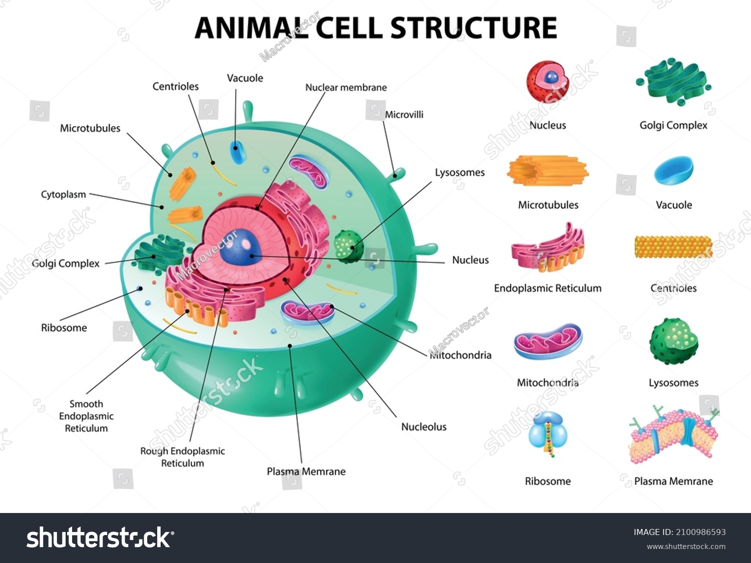
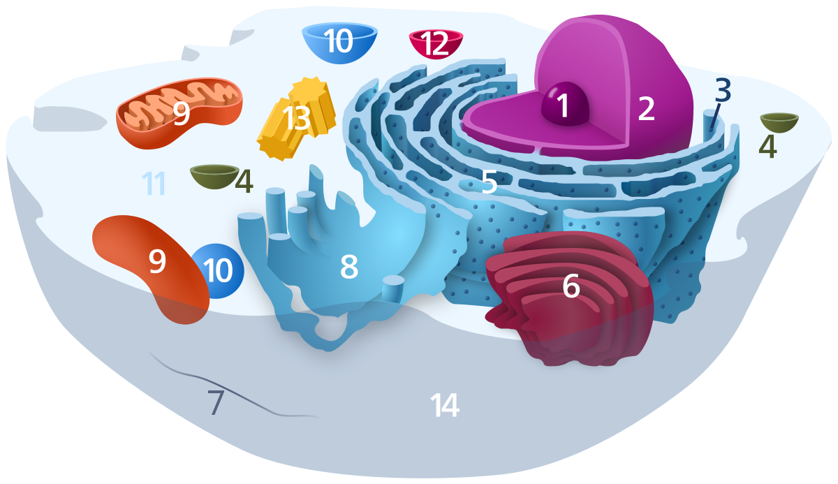
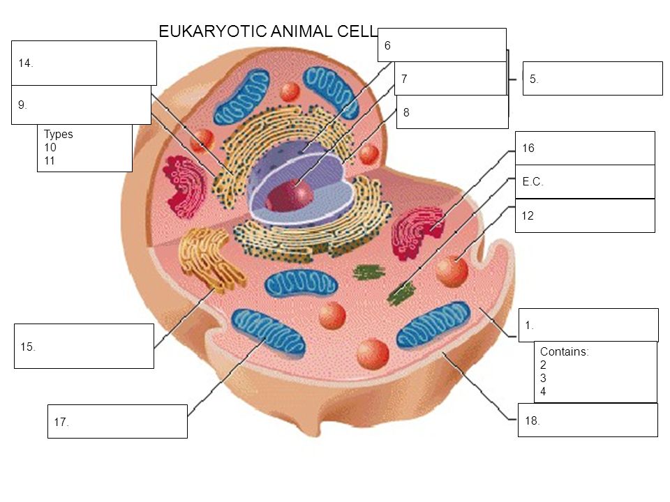
A Labeled Diagram of the Animal Cell and its Organelles A Labeled Diagram of the Animal Cell and its Organelles There are two types of cells - Prokaryotic and Eucaryotic. Eukaryotic cells are larger, more complex, and have evolved more recently than prokaryotes. Where, prokaryotes are just bacteria and archaea, eukaryotes are literally everything else.
Plant Cell - Definition, Structure, Function, Diagram & Types - BYJUS It is a rigid layer which is composed of polysaccharides cellulose, pectin and hemicellulose. It is located outside the cell membrane. It also comprises glycoproteins and polymers such as lignin, cutin, or suberin. The primary function of the cell wall is to protect and provide structural support to the cell.
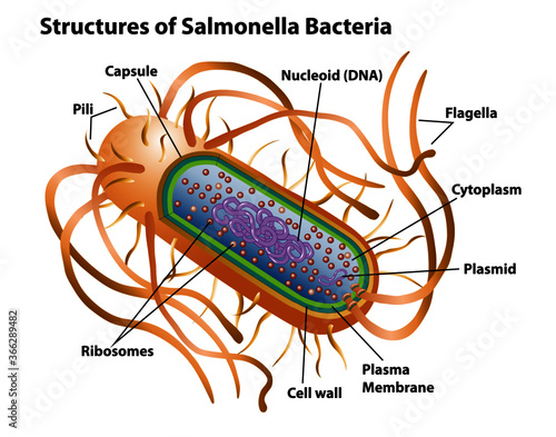
Bacteria Cell Structures with labels - Dreamstime Bacteria Cell Structures with labels Royalty-Free Vector Bacterial cell structures labeled on a bacillus cell with nucleoid DNA and ribosomes. External structures include the capsule, pili, and flagellum. Morphology of internal structures of bacteria. cell anatomy bacteria, prokaryotic cell, cell, internal structures, prokaryotic, dna, bacteria,
Structure of Fungal Cell (With Diagram) | Fungi - Biology Discussion The basic structural constituent of the cell wall in the Zygomycetes and higher fungi (Ascomycetes and Basidiomycetes) is chitin. It is a polysaccharide based on the nitrogen containing sugar (glucosamine). It is probable that more or less closely associated with chitin in the cell wall are pectic materials, protein, lipids, cellulose, callose ...
Cell Structure Quiz - PurposeGames.com Label the parts of the human cell. Label the parts of the human cell. English en. Login. Login Register Free Help; Start; Explore. Games; Playlists; Tournaments; Tags; The Wall; Badges; Leaderboard; ... This is an online quiz called Cell Structure. There is a printable worksheet available for download here so you can take the quiz with pen and ...
› cells › bactcellInteractive Bacteria Cell Model - CELLS alive Bacteria (Prokaryotes) are simple in structure, with no recognizable organelles. They have an outer cell wall that gives them shape. Just under the rigid cell wall is the more fluid cell membrane. The cytoplasm enclosed within the cell membrane does not exhibit much structure when viewed by electron microscopy.
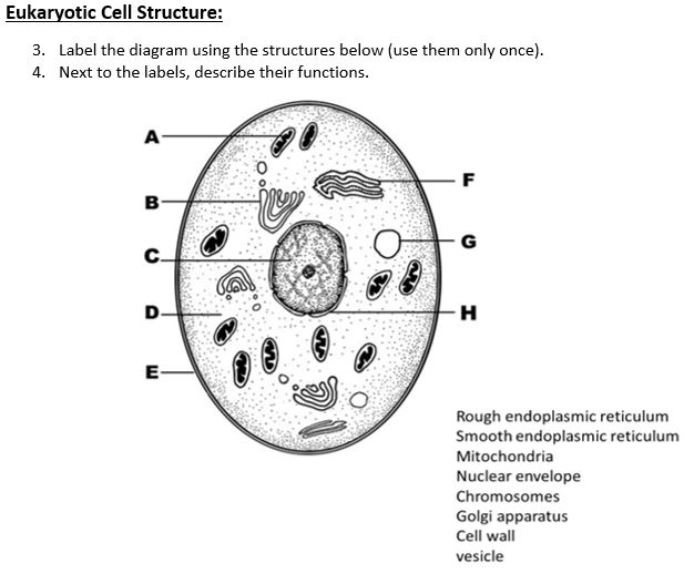
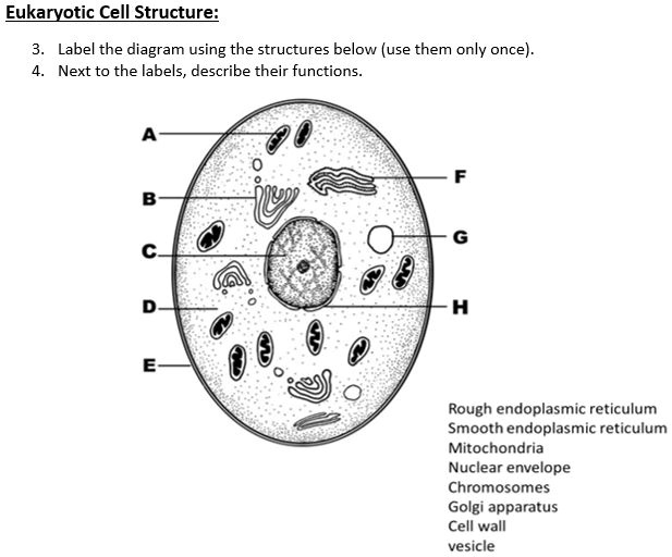
Eukaryotic Cell: Definition, Structure & Function (with Analogy ... Eukaryotic cells include animal cells - including human cells - plant cells, fungal cells and algae. Eukaryotic cells are characterized by a membrane-bound nucleus. That's distinct from prokaryotic cells, which have a nucleoid - a region that's dense with cellular DNA - but don't actually have a separate membrane-bound compartment like the nucleus.
Animal Cells: Labelled Diagram, Definitions, and Structure - Research Tweet Cilia and Flagella. Some eukaryotic cells either have cilia or flagella. Cilia are small, wiggling arm-like structures, whereas flagella are like a tail. Both structures are made of long protein fibers called microtubules, with a structure where nine microtubules form a ring around two central microtubules.
Label the cell structure. | Homework.Study.com Label the cell structure. Cells: All living cells contain an intracellular space called the cytoplasm. The cytoplasm is filled with a jelly-like fluid where many of the cells enzymatic reactions...
Plant Cell: Meaning, Components, Structure, Functions & Parts - Embibe Let us have a detailed look at the plant cell, its structure and the functions of different organelles. Components of a Plant Cell. The small membrane or non-membrane bound structures that are found in the cytoplasm or cellular matrix of a cell that works in a coordinated manner to maintain the homeostasis of a cell are termed as cell organelles.
Anatomy & Physiology Cell Structure & Function Quiz - Registered Nurse RN This Anatomy & Physiology (A&P) quiz is designed to test your knowledge of the basic cell structure and function. You will be asked questions that pertain to the mitochondria, nucleolus, nuclear membrane, ribosomes, lysosome, and much more. This practice test for the cell function and structure for Anatomy & Physiology, is designed to help you ...
en.wikipedia.org › wiki › CarmineCarmine - Wikipedia Carmine (/ ˈ k ɑːr m ə n, ˈ k ɑːr m aɪ n /) – also called cochineal (when it is extracted from the cochineal insect), cochineal extract, crimson lake, or carmine lake – is a pigment of a bright-red color obtained from the aluminium complex derived from carminic acid.









![1.1: Structure of the generalized cell [27]. | Download ...](https://www.researchgate.net/publication/325897026/figure/fig1/AS:639921136091136@1529580496082/1-Structure-of-the-generalized-cell-27.png)





![Cell structure - Stock Illustration [81732001] - PIXTA](https://t.pimg.jp/081/732/001/1/81732001.jpg)




Post a Comment for "42 cell structure with labels"